New advances in medical imaging technologies could significantly revolutionize existing clinical practices by enabling non-invasive and low-radiation alternatives for diagnostic purposes, according to speakers at a Thursday afternoon session of the 1996 Joint APS/AAPT Meeting in Indianapolis, Indiana. Sponsored by the APS Division of Nuclear Physics and the Forum on Industrial and Applied Physics, the session explored such emerging technologies as the application of positron emission tomography to laboratory testing of animals, a functional mammography technique using radiotracer methods to detect breast cancers, and digital x-ray imaging with active-matrix, flat-panel imagers.

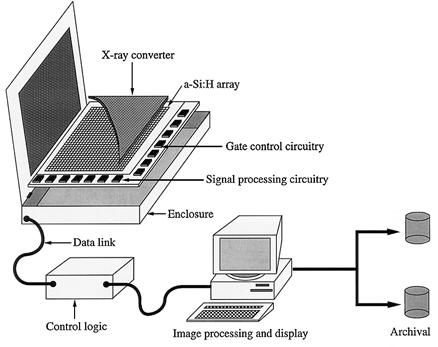
Schematic of active-matrix system
PETs for Imaging Laboratory Animals. Simon Cherry of the University of California, Los Angeles, described a positron emission tomography (PET) scanner that can detect features as small as 1.7 mm, compared to the four mm possible in conventional units.
In contrast to other medical imaging techniques, such as MRI or CAT scans, PET provides unique information on the rate of functional processes in the body. According to Cherry, one of its major strengths is the availability of positon-emitting isotopes of elements, such as carbon, nitrogen, oxygen, and fluorine. Many hundreds of compounds of these elements have been synthesized without changing their biochemical properties. By choosing the appropriate positon-emitting compound, PET can be used to measure biological processes such as blood flow, glucose metabolism, neutrotransmitter biochemistry, receptor affinity, enzyme activity, and gene expression.
However, while the process has been used clinically in the diagnosis and treatment planning of patients suffering from heart disease, cancer, epilepsy and Alzheimer's disease, its distribution to date has been limited to major research institutions and medical centers because of the high cost. There are currently fewer than 100 PET centers in the U.S. Many of these use the process as a biomedical research tool in animal studies.
Cherry's laboratory, in conjunction with CTI PET Systems in Knoxville, Tennessee, are developing MICROPET, the world's highest resolution PET for the imaging of small laboratory images. "This development opens the possibility of performing animal drug trials without the need for vivisection, saving animal lives and cutting costs for drug companies," said Cherry.
A use of MICROPET is to look at the functional effects of substance abuse in the monkey brain and to relate this to human drug addiction and its treatment. Other potential applications include drug testing, the effects of cancer treatments, and developmental studies in the normal and abnormal brain.
Still other PET-based technologies under development include a low-cost PET system to fit inside a conventional MRI machine to permit simultaneous PET and MRI imaging, providing images of the anatomy and function in the same imaging session. Also in the works is a low-cost detector system for imaging the axillary nodes in patients with suspected breast cancer, often the first site where cancer spreads.
Functional Mammography. In efforts to fight breast cancer, physicians employ mammography techniques - such as ultrasound imaging, magnetic resonance imaging, PET, and single-photon imaging with a gamma camera.
In efforts to improve detection of breast cancers, Irving Weinberg of PEM Technologies in Bethesda, Maryland described a complementary approach called functional mammography, a procedure that provides information on the biochemical processes in the breast by introducing radioactive tracers into the breast via the bloodstream. The tracers are then are imaged with a detector similar to those used in PET scans.
The detector system can be added to traditional x-ray mammography machines as a low-cost add-on, anticipated to be under $50,000. Unlike the gamma and PET cameras employed, which imaged areas of the body, functional mammography uses a detector system designed specifically to be placed on the breast, resulting in highly efficient signal detection. Weinberg hopes that this higher efficiency can be used to find smaller tumors, or reduce the patient radiation dose used in conventional nuclear medicine instruments.
Active-Matrix, Flat-Panel Imagers. Larry Antonuk of the University of Michigan in Ann Arbor described a pair of flat- panel imaging systems for diagnostic x-ray and cancer therapy imaging with image quality comparable to that possible through conventional techniques. Developed in collaboration between the University of Michigan and Xerox, these devices are capable of forming high-quality x-ray images in real time - as fast as 30 times per second.
As large as 26 centimeters on a side and a millimeter thick, the imaging circuits consist of thin-film transistors (TFTs) and photosensors made of amorphous silicon. Together these constitute an imaging pixel, with the TFT acting as a switch to control readout of signal generated in the photodiode. The array is covered by a phosphorescent material which serves to convert incident X rays to light.
According to Antonuk, flat-panel imagers offer a number of advantages. They are inherently resistant against radiation damage, so performance will not degrade in the high radiation environment of cancer therapy applications. Good x-ray sensitivity, low noise and large dynamic range offer the potential of superior image quality at reduced exposure to the patient. Finally, digital images allow quick processing on a computer or shared electronically. Other advantages include the compact, thin profile of the imagers, real-time acquisition and display, and a high linearity of response.
"There is every indication that the future imaging performance of optimized active-matrix, flat-panel imagers could meet or exceed that of existing radiographic, fluoroscopic, and portal imaging systems," said Antonuk.
©1995 - 2024, AMERICAN PHYSICAL SOCIETY
APS encourages the redistribution of the materials included in this newspaper provided that attribution to the source is noted and the materials are not truncated or changed.
July 1996 (Volume 5, Number 7)
Articles in this Issue

